Newsletter 2022.2 Index
Theme :"The Conference of Fluid Engineering Division (February issue)"
|
Stress of needle-free injection by focused microjet and study on stability of the jet velocity using machine learning
|
Abstract
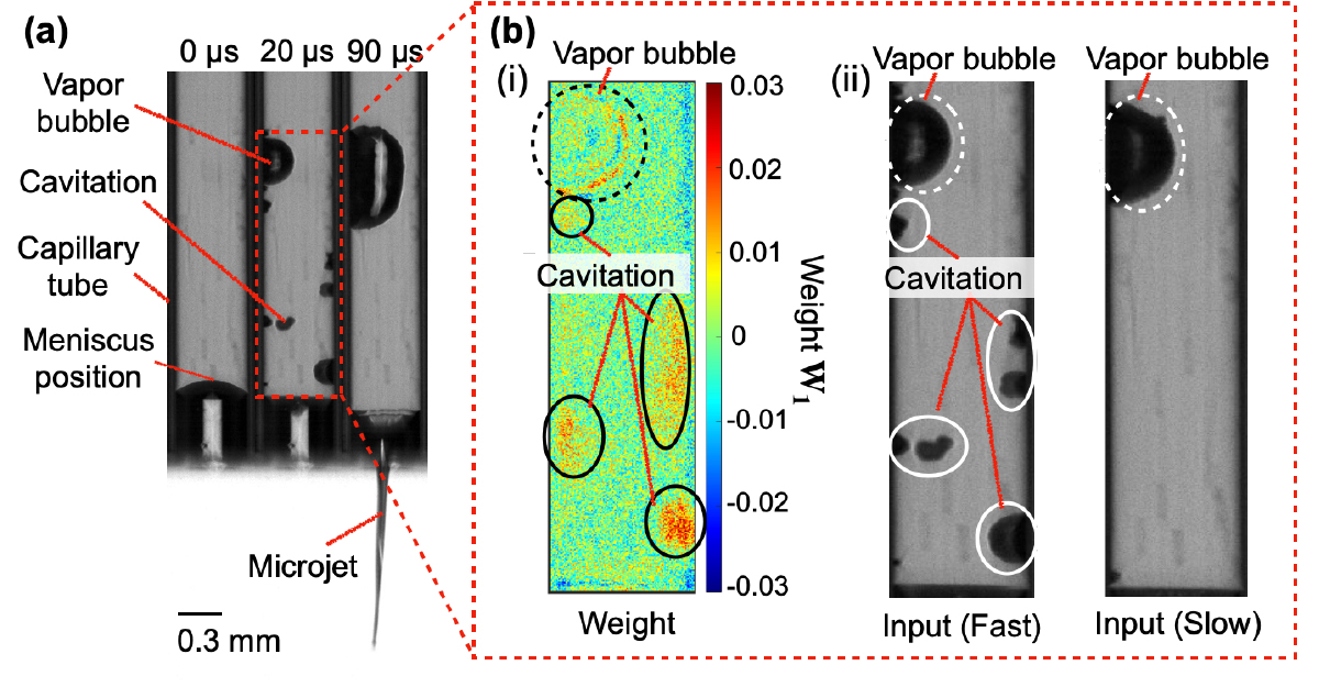
In this study, we visualized the stress field in human tissue simulant during penetration of needle-free injections of a highly focused microjet and a non-focused microjet(1). We also investigated the interaction between these injection methods and human tissue simulant by evaluating the dynamics of the induced cavity. We measured temporal evolution of cavity shape, stress intensity distribution, and stress vector field by using a high-speed polarization camera. As for the results, the cavity induced by focused microjet was dominated by shear stress and penetrated deep into tissue simulant with stress intensity lower than that by non-focused microjet. In contrast, the cavity induced by non-focused microjet was dominated by compressive stress in high stress intensity and the penetration depth was not as deep as that of a focused microjet. Such results indicate that the jet shape of focused microjet makes it advantageous for the development of minimally invasive medical devices. As the next step for the development of needle-free injection device with the focused microjet, the mechanism of the jet is being investigated with the aid of machine learning. We investigated the important features of laser-induced microjet images that affect the jet velocity by visualizing the image classification process of microjet images by a Feedforward Neural Network (FNN). The visualization of the trained weights suggested that the vapor bubbles and cavitation generated by the laser affect the jet velocity. Furthermore, cavitation has a larger effect on the jet velocity than vapor bubbles. Therefore, this study suggests that the air content in the liquid affects the stability of the jet velocity.
Key words
needle-free injector, microjet, polarization, stress visualization, machine learning, weight visualization
Figures

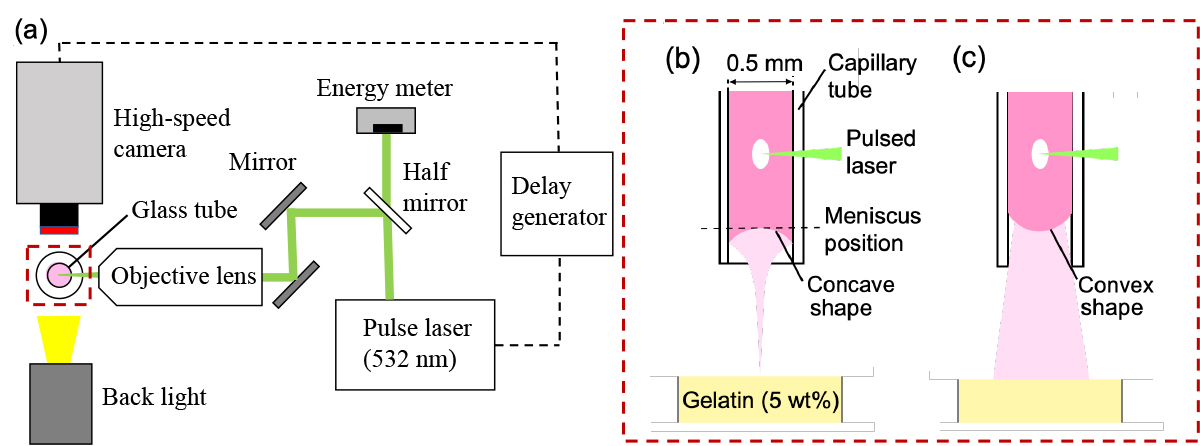
Figure 1 (a) Schematic diagram of experimental setup for the photoelastic measurement of stress field induced by (b) focused microjet, (c) non-focused microjet.

Figure 2 (a) Image sequence of stress fields and (b) stress vector fields induced by the penetration of(i) the focused microjet and (ii) the non- focused microjet.

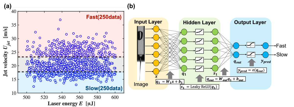
Figure 3 (a) Jet velocity Ujet vs. laser energy E in the experiment and (b) architecture of the FNN used in this study.

Figure 4 (a) Image sequence of the laser-induced microjet injection and (b) colormap of weight W1for classification of initial bubbles.
References


